Exploring the Microscopic World: Thermo Fisher Scientific Microscopy Imaging Contest 2024

By Sierra McConnell
The Thermo Fisher Scientific Microscopy Imaging Contest showcased some of the most stunning and scientifically significant microscopic images from researchers around the globe.
Entries were judged on four criteria, with the most important one being about the uniqueness and colorfulness of the submission. The full criteria were:
- Up to 5 points for using specific equipment
- Up to 10 points for using specific reagents and accessories
- Up to 2 points for using specific secondary antibodies and dyes
- Up to 30 points for showing multiple colors and unique shapes
The 2024 winners provided a fascinating glimpse into the intricate and often beautiful world of microscopic science. Let’s dive into some of the images from this year's contest.
Awesome Microscopic Images

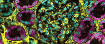
Studying SHH in Kidney Tissue
Lindsey Avery Fitzsimons, PhD
Why It’s Awesome: Imagine seeing the tiny parts of your kidney that help it function. This image shows a part called the primary cilium, which acts like a little antenna for the cell. Dr. Fitzsimons started studying this because she was diagnosed with a rare kidney disease. Her research could help doctors better understand and treat kidney problems, including her own.

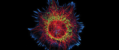
The Cytoskeleton of a Neuroblastoma Cell
Bruno Cisterna, PhD
Why It’s Awesome: This image is like a colorful blueprint of a mouse neuroblastoma cell, which is a type of cancer found in nerve cells. You can see different parts like microtubules and mitochondria, which are crucial for the cell’s functions. Dr. Cisterna’s research helps us understand how the cell’s structure affects the movement of essential materials inside it. It’s like figuring out how the roads and highways inside a city work!

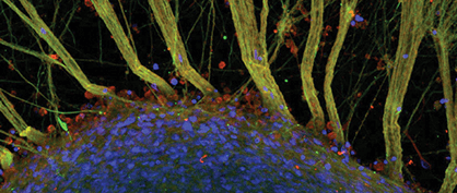
Human Neural Networks
Stuart Hodgetts, PhD
Why It’s Awesome: Have you ever wondered what the inner workings of your brain look like? This image shows the intricate network of neural fibers in the human brain. It looks like a beautiful web of connections, all ready to help you complete different thoughts or actions. Dr. Hodgetts is working on ways to repair spinal cord injuries and help nerves regenerate. His research could help people recover from serious injuries and regain their abilities.
These images not only highlight the beauty of scientific exploration but also underscore the profound impact of research in advancing our understanding of complex biological systems. The Thermo Fisher Scientific Microscopy Imaging Contest continues to inspire and engage the scientific community, bridging the gap between art and science.
Check out the rest of the winners, more stunning images, and learn more about the contest here.